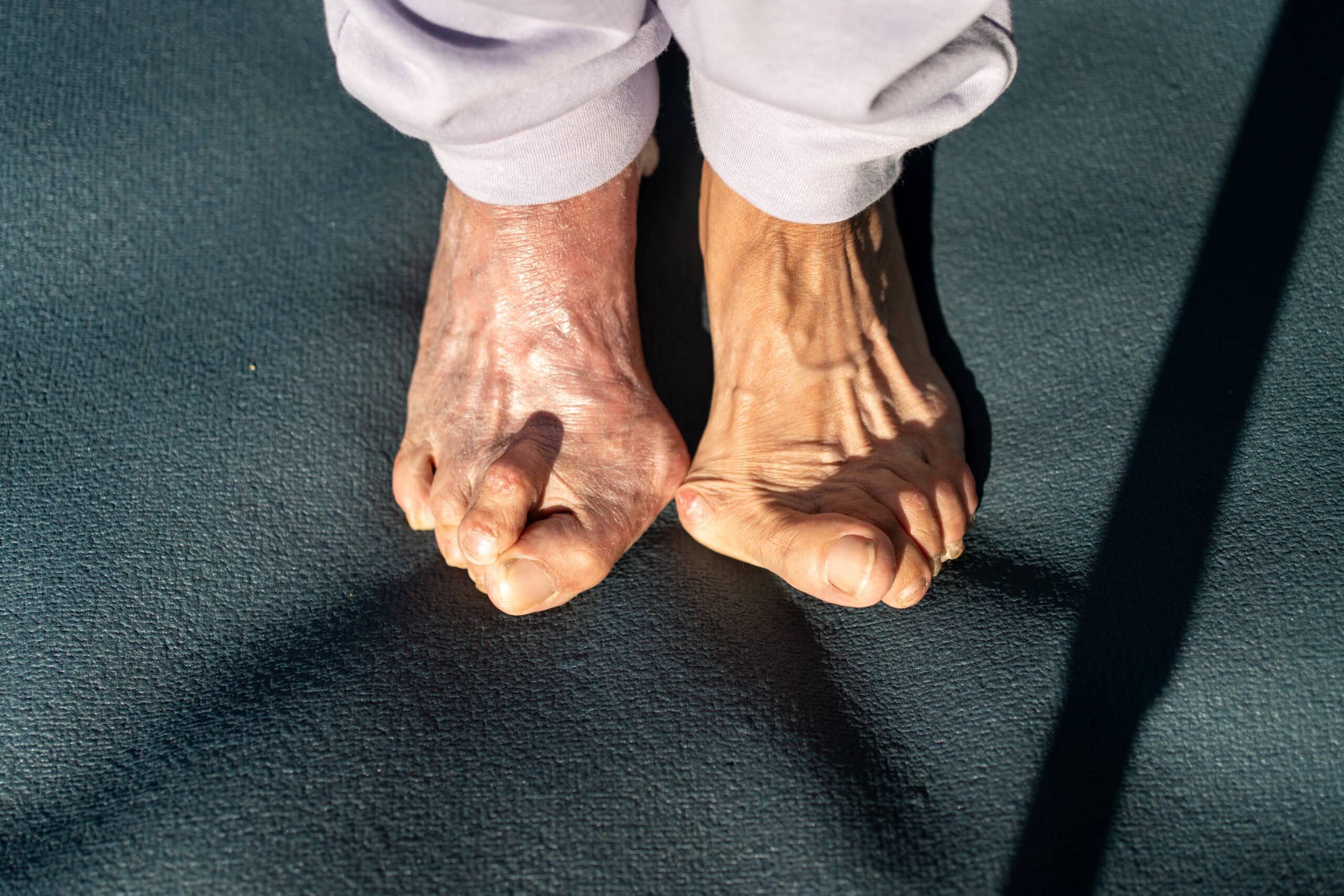
Big toe arthritis joint preservation versus fusion?
Arthritis rarely takes over a person’s life all at once. It usually negotiates its way in gradually. First, a certain shoe stops working. Then longer walks become less appealing. Exercise changes. Workdays become harder. Eventually, patients can find themselves planning ordinary parts of life around one painful joint.
By the time many patients come to see me, they have already been told some version of the same story: there is arthritis in the joint, a bone spur has formed, and the eventual answer may be fusion. Sometimes fusion is the right answer. But an arthritic joint on an X-ray does not automatically tell me whether the joint must be sacrificed now, and a visible bone spur does not tell me that removing the spur alone will adequately treat the problem.
The more important questions are clinical. Where does the joint hurt during motion? Is the pain concentrated at the point where the toe jams against the spur, or is it present throughout the arc of motion? Is there grinding inside the joint? How much useful motion remains? What forces are continuing to compress the joint? And, most importantly, what does the patient need the joint to do in the life they want to keep living?
That is where toe arthritis joint preservation planning begins.
The short answer is that some arthritic big toe joints can still be preserved. The decision is not based on the X-ray alone, however, and preservation is not automatically the better choice. It depends on the pain pattern, the quality of the remaining motion, the forces compressing the joint, and whether that motion can still serve the patient’s actual goals.
Hallux limitus and hallux rigidus are not just X-ray diagnoses
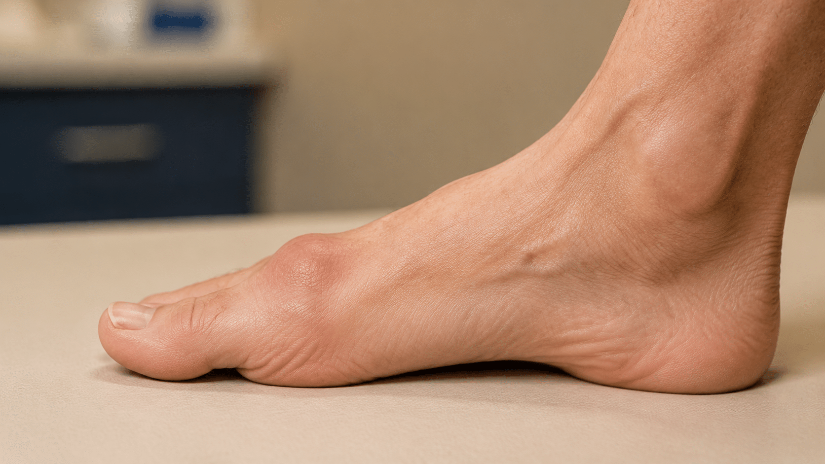
Arthritis at the base of the big toe is commonly described as hallux limitus when motion is restricted and as hallux rigidus when the loss of motion and arthritic disease are more advanced. The joint may develop cartilage wear, narrowing of the joint space, subchondral sclerosis, cystic changes, and bone spurs, especially along its upper surface.
Those findings matter, but they do not make the decision by themselves. Two patients with similar-looking radiographs can have very different pain patterns, functional limitations, remaining motion, and goals. One may feel a sharp block only when the toe reaches the end of its upward motion. Another may feel pain and grinding through nearly every part of the movement. Those are not the same clinical problem, even if both are labeled big toe arthritis.
This is why I do not begin with the question, “Which operation matches this grade of arthritis?” I begin by asking why the joint hurts and what useful function may still be preserved.
Where the pain occurs may matter as much as how the X-ray looks
One of the most useful distinctions is whether pain occurs primarily at the end of motion or throughout the motion.
Pain at the end of motion
When pain is concentrated at terminal dorsiflexion – the point at which the big toe bends upward as far as it can – the dorsal bone spur may be acting as a mechanical doorstop. The toe moves until bone strikes bone, producing a painful jamming sensation. This is the classic pattern that can respond well to cheilectomy, the removal of the obstructing bone.
Pain through the middle of motion
Pain in the middle of the joint’s motion, especially when accompanied by substantial crepitus or grinding, suggests more extensive articular disease. Instead of one painful collision at the top of the joint, the damaged surfaces may hurt as they move across one another. Joint-preserving surgery may still be discussed in carefully selected cases, but the expected result is less predictable and fusion or replacement becomes a more important part of the conversation.
Pain that is mechanically redistributed
The big toe joint is not only its dorsal surface. It also includes the plantar joint, sesamoid complex, capsule, tendons, and surrounding soft tissues. Changing one part of the system changes how other parts are loaded. That becomes especially important after cheilectomy.
Removing the spur is not always the same as treating the joint
A cheilectomy removes the dorsal osteophyte and opens the blocked portion of the joint. For the right patient, that may reduce impingement pain and improve motion without sacrificing the native joint. Published studies support cheilectomy as an established joint-preserving option, particularly when symptoms are concentrated near the end of motion.
But the spur is sometimes the most visible consequence of a deeper mechanical problem rather than the entire problem itself. If the joint remains compressed, simply clearing the top may create additional motion without creating enough functional space inside the joint.
That distinction is central to my approach. I am not only trying to make the toe move farther during an examination. I am trying to determine whether the joint can use that motion more comfortably during walking, standing, work, exercise, and the patient’s chosen footwear.
More motion is not automatically better motion. The goal is useful motion.
Useful motion is not an abstract number. It is the motion that allows the patient to move through a work shift, push off during a walk, manage uneven ground, return to an activity, or wear the shoes that fit the life they want to live.
The paradox of postoperative motion
It seems logical that if a painful joint is blocked, removing the block and gaining motion should always help. Often it does. But an arthritic joint can expose a paradox: the new motion must be absorbed by cartilage, the plantar joint, the sesamoids, the capsule, and soft tissues that may already be damaged, stiff, or irritated.
The dorsal impingement pain may improve while a deeper, plantar, or redistributed pain becomes more noticeable. This does not necessarily mean the cheilectomy was technically unsuccessful. It may mean that the procedure created motion the remaining joint was not fully prepared to use.
That possibility deserves an honest discussion before surgery. It is one reason I evaluate the pain pattern rather than relying on the spur alone, and one reason I may recommend meaningful joint decompression rather than an isolated cheilectomy.
Root: determine why this joint hurts
The first stage of the PRISM™ approach is identifying the root of the problem. That evaluation may include weight-bearing radiographs, hands-on examination of the joint through its available motion, assessment of the sesamoid region and plantar structures, and analysis of the mechanics affecting the first ray and forefoot.
I want to know whether the pain is primarily an impingement problem, a pressure problem throughout the joint, a broader mechanical problem, or some combination. I also want to understand the destination. A patient who stands for full retail shifts, a patient who wants to return to running or yoga, and a patient who primarily wants dependable walking comfort may define useful motion differently.
The role of the guide begins with understanding where we need to go. Without that destination, even a technically good operation can solve the wrong problem.
Fix: create functional space, not just a cleaner X-ray
When the native joint retains a reasonable opportunity to provide useful, comfortable motion, I try to preserve it. The exact correction is individualized, but it may combine thorough removal of the obstructing dorsal bone with a decompression osteotomy of the first metatarsal.
The osteotomy is conceptually related to a modified bunion-type bone cut, but its purpose here is different. It changes the relationship within the joint so that pressure can be reduced and functional space created. Instead of only clearing the roof of a crowded joint, the reconstruction addresses the crowding itself.
This approach is supported by a growing body of joint-preservation literature. Registry data have shown improvement after both cheilectomy and metatarsal decompression osteotomy in moderate hallux rigidus, with higher satisfaction in the osteotomy group in one national-registry study. A 2026 systematic review found that decompressive first-metatarsal osteotomy, usually performed with cheilectomy, may improve pain, function, and motion in selected patients with early or moderate disease. The authors were also appropriately cautious: most available studies are observational, cheilectomy was frequently performed at the same time, and patient selection remains critical.
That is consistent with how I use the procedure. It is not a universal alternative to fusion. It is a strategy for a joint that still has a reasonable chance to provide useful motion if the obstruction and compression are addressed together.
The minimally invasive technique matters here, but not because a small incision can substitute for a complete correction. Working through small portals requires specialized judgment about how much bone to remove, how to decompress the joint, and how to preserve the surrounding tissues while still accomplishing enough structurally. The goal is minimal disruption, not minimal correction.
Heal: the surgery and the healing environment are one plan
Structural correction is only one part of the outcome. The joint must then heal, adapt to its new space, and function within the mechanics of the rest of the foot.
PRISM™ is the surgeon-designed system through which I orchestrate that entire process. It is not an upgrade added to a conventional operation, a fixed package, or a collection of isolated treatments. The pillars are consistent, but their application and sequencing are individualized. I begin with the patient, the joint, the healing profile, and the outcome we are trying to reach, then coordinate every part of the correction and recovery around those needs.
For a toe arthritis joint preservation case, the plan may draw from a wide range of regenerative, recovery, stability, and mechanical strategies. The exact combination is selected by the surgeon according to the procedure, the condition of the joint, the patient’s biological and mechanical risk profile, and the demands of the life that patient wants to return to.
The objective is orchestration. The bone work, joint environment, stability, recovery progression, footwear, and long-term mechanics should not function as separate parts. They should be intentionally coordinated by the surgeon to support the same destination.
What regenerative support can – and cannot – do
An arthritic joint does not need perfect cartilage for joint-preserving surgery to provide meaningful benefit. Patients may have cartilage loss, narrowing, sclerosis, and other arthritic changes and still retain a usable joint. The harder question is whether enough functional capacity remains for preservation to make sense.
In my observed clinical experience, patients selected for thorough decompression often have a better pain and recovery trajectory when biologic support is incorporated into the same plan. Published clinical research has also explored amniotic tissue as an adjunct to cheilectomy, including a prospective randomized study. That adjunctive evidence remains limited. It does not establish that a biologic product will regrow normal cartilage, reverse advanced arthritis, or rescue a joint that is not mechanically preservable.
That is not the promise. The purpose is to support the healing environment of a joint we have already judged reasonably preservable. Biology cannot compensate for the wrong structural operation, and a technically excellent operation cannot make severely diseased cartilage normal.
How I decide whether a joint is reasonably preservable
No single finding answers the question. I look at the relationship among several factors:
whether pain is limited mainly to end-range impingement or occurs throughout motion;
the degree of crepitus and the quality of the remaining motion;
cartilage and joint-space changes on weight-bearing radiographs;
the anatomy and mechanics contributing to compression;
the condition of the plantar joint and sesamoid complex;
age, activity, bone quality, footwear expectations, and recovery priorities;
what tradeoffs the patient is willing to accept; and
whether the remaining joint appears capable of providing useful motion after decompression.
This is not a contest to preserve every joint at any cost. Preservation has value only when the likely function justifies its uncertainty.
Fusion is not a failure – but it is a permanent tradeoff
Fusion of the first metatarsophalangeal joint can be an excellent operation for end-stage arthritis. It can drastically reduce crippling joint pain and provide a stable platform for walking. I do not reject fusion, and I do not present it as an inferior or destructive treatment.
It is, however, motion-sacrificing by design. Once the joint is fused, its position must serve several competing needs: standing, gait, push-off, terrain, activity, and footwear. Other joints and structures must accommodate the motion that no longer occurs at the big toe joint. Many patients function very well after fusion, but a healed fusion does not make the foot completely normal, guarantee a pain-free foot, or create a mechanically perfect solution for every part of a person’s life.
My position is straightforward: fusion is not a procedure I reject. It is a permanent option I try not to use before its tradeoffs clearly make more sense than preserving motion.
When pain is present throughout the joint, crepitus is substantial, cartilage loss is advanced, or the remaining joint cannot reasonably be expected to use additional motion, fusion may provide a more dependable outcome than a preservation attempt. The point is not to delay fusion reflexively. The point is to choose it deliberately.
What about big toe joint replacement?
Joint replacement is another motion-preserving option for selected patients, but it brings its own considerations, including implant durability, bone quality, activity demands, anatomy, remaining motion, and the possibility of revision. It is not interchangeable with native-joint preservation and it is not automatically preferable to fusion.
Preserving the native joint when clinically reasonable can keep future options open, including replacement or fusion if arthritis progresses or preservation eventually fails. But future optionality should never be used to justify an operation that is unlikely to help now.
Why I reserve 90 minutes for this consultation
Toe arthritis joint preservation surgery requires more than assigning a procedure to an X-ray grade. It requires a real discussion about what the patient wants, what useful motion means in that person’s life, how much uncertainty is acceptable, and what each pathway can and cannot accomplish.
I reserve 90 minutes for an initial consultation because that is what this work requires. The time is not a luxury amenity. It is a clinical tool. It allows me to understand the patient’s destination, examine the joint and the mechanics around it, study the imaging, explain the possibility of a different postoperative pain pattern, and compare preservation, replacement, and fusion without forcing a permanent decision into a compressed encounter. The consultation is not a commitment to surgery, and preservation is not a predetermined recommendation. Its purpose is to make the correct decision carefully.
The right outcome starts with understanding why the problem happened in the first place. From there, we can design the right fix and the environment in which it has the best opportunity to heal.
Frequently asked questions
Does big toe arthritis always lead to fusion?
No. Some joints remain candidates for preservation, particularly when pain is concentrated at end-range impingement and useful motion remains. More advanced disease, pain throughout motion, and substantial crepitus make preservation less predictable and may make fusion more appropriate.
Is a cheilectomy just removal of the bone spur?
At its core, cheilectomy removes obstructing bone from the top of the joint. For some patients, that is enough. For others, the joint remains compressed and a decompression osteotomy may be considered to create more functional space.
Will surgery restore normal cartilage?
No operation or biologic treatment should be presented as a guarantee of normal cartilage restoration. The goal of joint preservation is to reduce the mechanical sources of pain, preserve useful native motion when reasonable, and support the joint’s healing environment.
Does damaged cartilage automatically rule out joint preservation?
Not necessarily. Cartilage does not have to be perfect for a joint-preserving procedure to help, but the extent and location of the damage matter. Pain throughout motion, substantial crepitus, global joint-space loss, and poor remaining function make preservation less predictable. The question is whether the joint retains enough biological and mechanical capacity to provide useful motion after decompression.
Can joint-preserving surgery fail?
Yes. Arthritis surgery contains uncertainty. Pain can persist, a different pain pattern can emerge, arthritis can progress, and later replacement or fusion may become necessary. Careful selection and counseling reduce surprises but cannot eliminate risk.
Why not choose fusion immediately if it is reliable?
Fusion may be the best choice for an end-stage, globally painful joint. In a reasonably preservable joint, however, it permanently removes motion and changes how the foot accommodates gait, footwear, and activity. The decision depends on which tradeoff best fits the patient.
Is PRISM™ the same for every patient?
No. PRISM™ is not a standardized bundle. It is the surgeon-led orchestration of correction, regenerative support, healing optimization, stability, mechanical protection, and recovery progression. Each element is individualized for the patient, the procedure, and the outcome being pursued, then coordinated as one complete plan.
A real example of what joint preservation can mean
One of my patients had spent more than ten years unable to wear many of the shoes she owned. Her big toe arthritis affected full retail shifts and even what she felt able to wear to church. Her joint-preservation plan addressed the obstructing bone, the joint compression, the healing environment, and the mechanics that would protect the result. Her early recovery was more painful than most patients experience in my practice, but we treated the pain and stayed closely involved. She ultimately regained strong motion, returned to regular shoes, and told me, “I’m now able to fit into shoes I haven’t worn in over 10 years.”
Schedule a comprehensive surgical consultation
If big toe arthritis is limiting your work, walking, activity, or footwear, the first question is not simply whether an X-ray looks severe. The first question is whether the joint still has a reasonable opportunity to provide useful, comfortable motion – and which tradeoffs make sense for your life.
Dr. Michael Tagge’s initial surgical consultation is a 90-minute, surgeon-led evaluation. The practice is cash-pay and out of network so the evaluation and plan can be built around what best solves the problem, including clinically purposeful elements that may fall outside conventional insurance pathways.
Medical note
This article provides general educational information and does not establish a diagnosis or recommend a specific procedure. Candidacy, recovery, risks, and results vary by patient.
Clinical references